The PPG hardware delay and the adopted pulse arrival time
VitalDB's raw ECG→PPG pulse arrival time is about 0.4 s longer than physiology. Splitting it into a hardware delay (one value for the release; One device delay for the PLETH across the release) and a physiological part needs an adopted value from studies that sampled ECG and PPG on one clock: about 270 ms for anaesthetised adults.
| Date | 2026-10-12 |
| Status | current |
| Methods | Delays and averaging windows, Reading the source |
| Clocks | Pulse oximeter frame clock |
| Devices | Solar8000 monitor, SNUADC |
| Variables | Plethysmogram (SNUADC), ECG lead II (SNUADC) |
Background
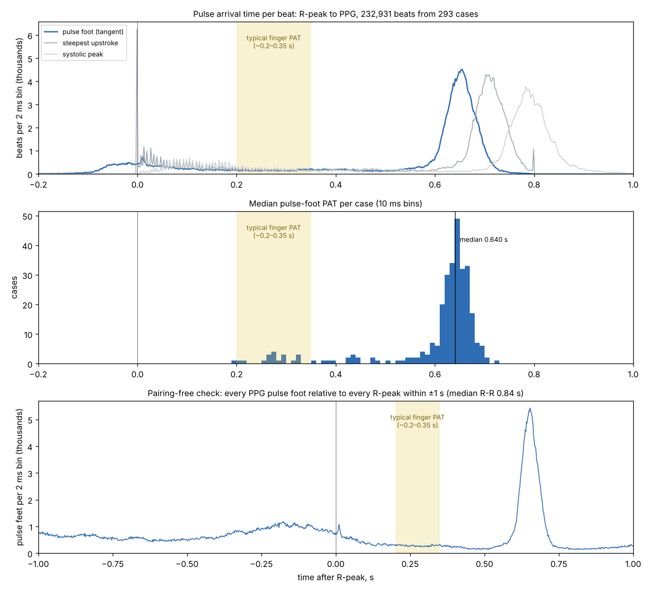
The median R-to-PPG-foot time in VitalDB is 0.64 s, against roughly 0.2–0.3 s in adults measured with shared-clock hardware; an earlier VitalDB study found 645 ± 26 ms. The excess is the monitor's buffering and filtering, and it cannot be separated from physiology inside VitalDB.
Data
232,931 beats in 293 cases (raw PAT); published shared-clock studies of awake adults.
Method
Literature graded by how ECG and PPG were clocked: A (one acquisition unit), B (research equipment, shared clock likely), C (clinical monitor, excluded). Adults with an R-peak ECG fiducial and finger PPG foot.
Results
- Tier A studies put R-to-finger-foot PAT at about 200–285 ms in awake adults; none gives a verifiable shared-clock value under anaesthesia.
- Anaesthesia lengthens PAT by about 40 ms in monitor-based work (240 ms before induction, 279–284 ms after).
- For VitalDB (adults under general anaesthesia, mean age about 58): about 270 ms, plausible range 250–290 ms.
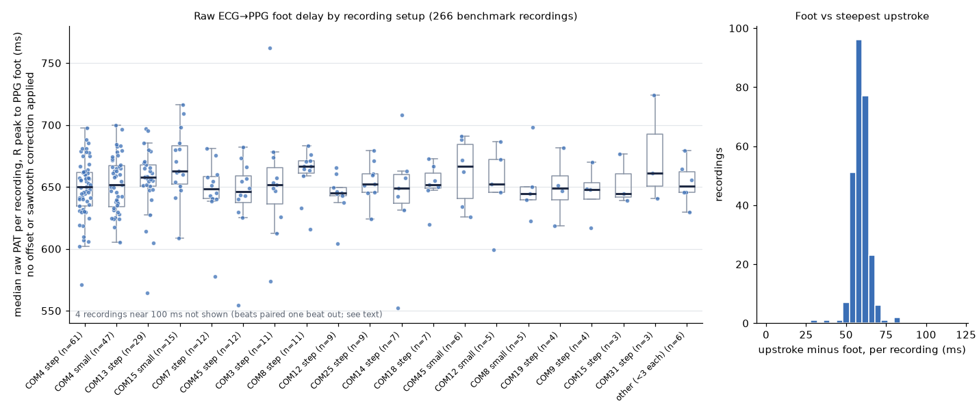
- Hardware delay with that value: about 385 ms over these 293 cases (to the PPG foot). Per-setup values of 368–399 ms were reported here first; the full census (One device delay for the PLETH across the release, 5,536 recordings) found no difference between setups beyond ±15 ms and the same value, 385 ms (655 ms to the foot minus 270 ms), so one global delay is used. The adopted value and the delay must use the same fiducial: 270 ms is to the foot.
| Parameter | Value | Status | From |
|---|---|---|---|
| Adopted mean pulse arrival time | 270 ms (plausible range 250–290 ms) | assumed | The PPG hardware delay and the adopted pulse arrival time |
Discussion
The adopted value must use the same PPG point the software detects. The global delay is taken at the foot, where the 270 ms applies; a PAT timed to the steepest upstroke comes about 60 ms later, and that difference stays in the corrected value (Pulse arrival time in two correction steps). Since the census, one delay serves every recording, so a change to the adopted value moves every recording's level by the same amount.
Observations it supports
| Observation | Status | Summary |
|---|---|---|
| The PLETH arrives about 0.4 s late | established | Raw R-to-PPG-foot pulse arrival time in VitalDB has a median of 0.64–0.71 s, about 0.4 s more than physiology: the monitor's buffering and filtering. One delay fits the whole release: no clusters by setup, SNUADC port or case order (One device delay for the PLETH across the release). |
| The plethysmogram is built in 30 Hz frames | established | The SNUADC PLETH waveform comes from the oximeter in 33.3 ms frames, smoothed to 500 Hz. Its content changes once per frame, on the oximeter's own clock, about 0.4 s after the finger. |
Limitations
The hardware delay is not observable in VitalDB; it rests on the adopted physiological value.
Worked examples
| Example | Side | Status | Summary |
|---|---|---|---|
| Pulse arrival time against blood pressure, before and after synchronisation | time | draft | In 3,270 recordings, PAT tracks systolic pressure only once each pulse is paired with its own beat: the hourly correlation moves from a mean of −0.04 to −0.21 (88% of recordings negative), reproducing Ruffolo et al. 2025 in a second dataset; paired naively, PAT is a heart-rate signal. |
| Pulse arrival time in two correction steps | time | draft | The time from the ECG R wave to the pulse at the finger, as stored, after the PLETH's sawtooth is removed, and after its device offset is removed, smoothed by maximum likelihood, with the arterial line as the reference. |
References
- Allen J, Murray A. Variability of photoplethysmography peripheral pulse measurements at the ears, thumbs and toes. IEE Proc Sci Meas Technol. 2000;147(6):403-407. ↗
- Allen J, Murray A. Age-related changes in peripheral pulse timing characteristics at the ears, fingers and toes. J Hum Hypertens. 2002;16(10):711-717. ↗
- Block RC, Yavarimanesh M, Natarajan K, et al. Conventional pulse transit times as markers of blood pressure changes in humans. Sci Rep. 2020;10:16373. ↗
- Jose AS, Srivastav S, Mehta B. Exploring the repeatability of pulse arrival time in healthy subjects: a test-retest approach. Indian J Physiol Pharmacol. 2025;69(1):27-33. ↗
- Kortekaas MC, van Velzen MHN, Grüne F, Niehof SP, Stolker RJ, Huygen FJPM. Small intra-individual variability of the pre-ejection period justifies the use of pulse transit time as approximation of the vascular transit. PLoS ONE. 2018;13(10):e0204105. ↗
- Lee J, Yang S, Lee S, Kim HC. Analysis of pulse arrival time as an indicator of blood pressure in a large surgical biosignal database: recommendations for developing ubiquitous blood pressure monitoring methods. J Clin Med. 2019;8(11):1773. ↗
- Liao S, Liu H, Lin W-H, Zheng D, Chen F. Filtering-induced changes of pulse transmit time across different ages: a neglected concern in photoplethysmography-based cuffless blood pressure measurement. Front Physiol. 2023;14:1172150. ↗
- Martin SL-O, Carek AM, Kim C-S, Ashouri H, Inan OT, Hahn J-O, Mukkamala R. Weighing scale-based pulse transit time is a superior marker of blood pressure than conventional pulse arrival time. Sci Rep. 2016;6:39273. ↗
- Mohammadpoor Faskhodi M, Garcia-Gonzalez MA, Fernandez-Chimeno M, et al. On the use of fractional calculus to improve the pulse arrival time (PAT) detection when using photoplethysmography (PPG) and electrocardiography (ECG) signals. PLoS ONE. 2024;19(2):e0298354. ↗
- Obata Y, Ong QJ, Magruder JT, et al. Noninvasive assessment of the effect of position and exercise on pulse arrival to peripheral vascular beds in healthy volunteers. Front Physiol. 2017;8:47. ↗
- van Velzen MHN, Loeve AJ, Niehof SP, Mik EG. Increasing accuracy of pulse transit time measurements by automated elimination of distorted photoplethysmography waves. Med Biol Eng Comput. 2017;55(11):1989-2000. ↗
- Youn AM, Shin YS, Park SI. Changes in pulse transit time according to target controlled infusion of propofol versus sevoflurane inhalation induction. Anesth Pain Med. 2014;9(1):48-53. ↗
- Ruffolo I, Siddiqui A, Nguyen B, Dixon W, Assadi A, Greer R, Schwartz S, Brudno M, Mariakakis A, Goodwin A. High-fidelity measurement of pulse arrival time in critically ill children using standard bedside monitoring equipment. Physiol Meas. 2025;46(11):115006. ↗
Figures