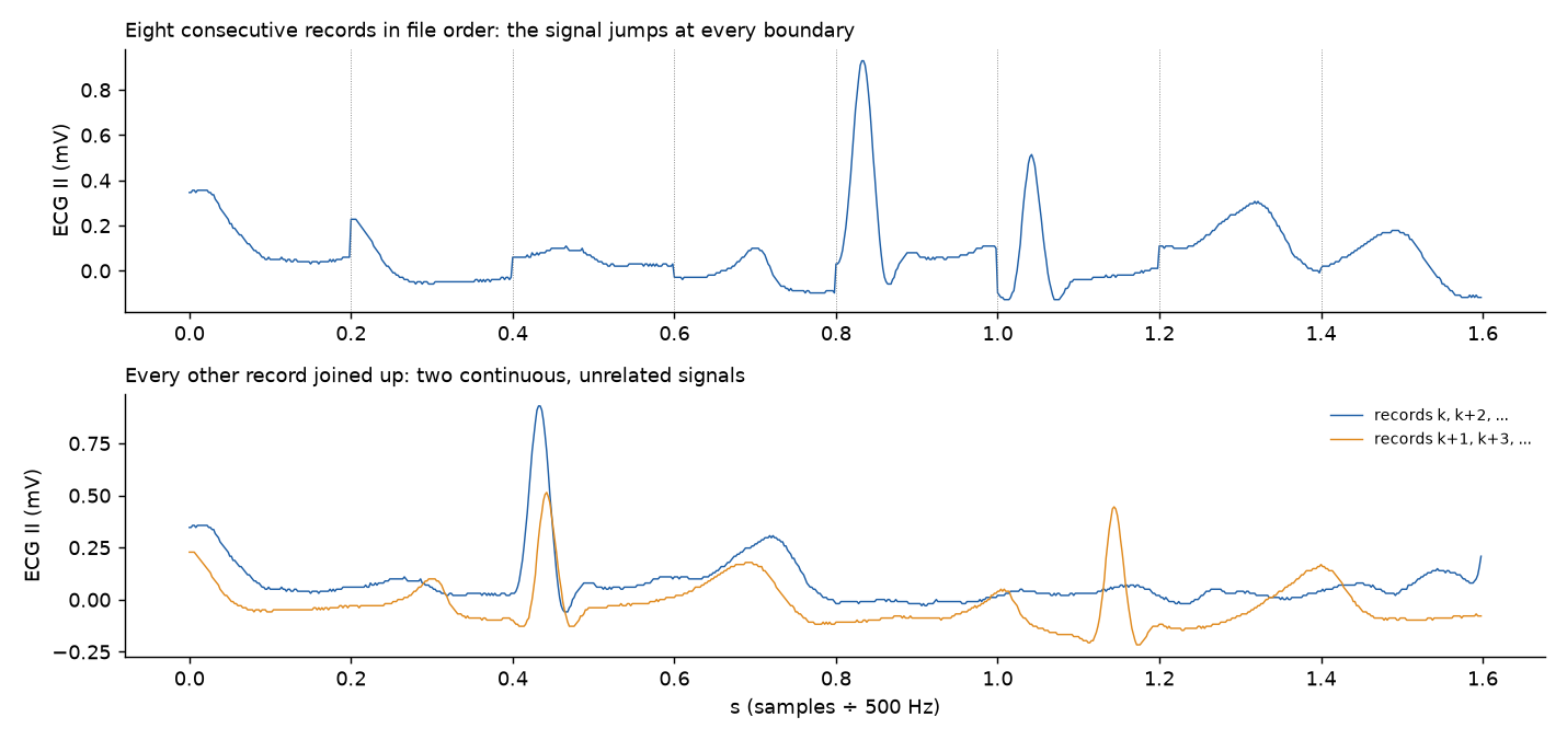
In some recordings two unrelated streams are interleaved in one track
In 7 recordings 64–95% of the records alternate between two streams: each record continues the one two before it, not the one before, and the two streams do not match each other. SNUADC and BIS in case 2219, case 2379, case 2640, case 4836, case 5327 and case 5617; SNUADC in case 1939. In case 94, case 945, case 1132, case 3195 and case 5700 4–7% of SNUADC records interleave.
| Status | open: seen, not explained. |
| Devices | Vital Recorder, SNUADC, BIS monitor |
| Variables | ECG lead II (SNUADC), Arterial pressure (SNUADC), Plethysmogram (SNUADC), BIS EEG |
| Supersedes | In 34 files the SNUADC records overlap throughout |
What it means
The timestamps alternate (13 / 187 ms or 23 / 177 ms between SNUADC records, pairs summing to one 0.2 s message), and the file holds about twice as many samples as its time span allows. Read in file order, the track switches between two signals every 0.2 s. Joining each record to the one two before gives two continuous signals (boundary jump 0 codes, against 11 to the neighbouring record); their correlation is 0.10 and 0.01 in the two cases tested, with different amplitudes, so they are not two copies of one signal. ECG II, ART and PLETH interleave together, and BIS does too, so the recorder merged two sources. Which stream belongs to the patient is not known; in one case one stream's rhythm matches the monitor's heart rate better.
What is done about it
Treat these tracks as unusable until the streams are separated and attributed. A single run of backward steps that pair to one message marks an interleaved stretch.
Evidence
| Study | Date | Status | Summary |
|---|---|---|---|
| Message sizes and the steps between records | 2026-10-19 | draft | Every record in every waveform: SNUADC writes messages of 100 samples (0.2 s), Primus 30 (0.48 s), BIS 16 (0.125 s), CardioQ no fixed size. Backward steps outnumber gaps by about a hundred to one and come from a few recordings with duplicated or interleaved records. Primus gaps are whole lost messages; most SNUADC and BIS gaps are not. |
Figures